The RETAIN project performed teleradiology, with synchronization and videophony allowing teleworking, over the European International Broadband Communications (IBC) network prototype. Trials were between radiologists at an European scale. The network was the Pan-European ATM pilot. The project was supported by a grant of the EU, TEN-IBC programme.
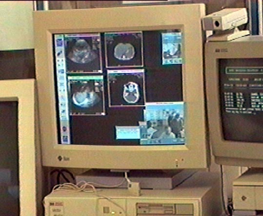
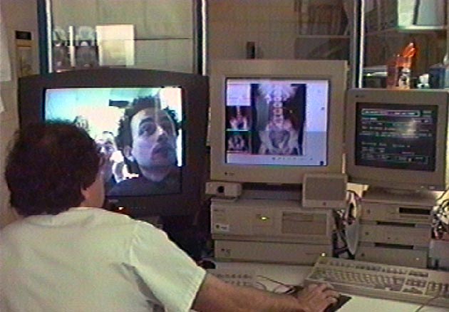
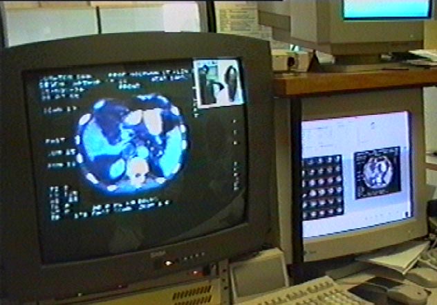
The test system is shown below.
 |
 |
 |
|
RETAIN - Phase 2
|
RETAIN - Phase 3
|
RETAIN - Phase 3
|
Other Retain Web sites:
Phase II elapsed from November 1994 to April 1995, TEN-IBC B2012 project, performing IBC Trials between radiologists at an European scale: Rennes (F), Barcelona (E),and Oldenbrug (D), over the European ATM Pilot network.
Phase III, elapsing from November'95 to November'96, will extend sessions to Coimbra (P), Duesseldorf (D),and Paris (F), and will try to run multiconference IBC sessions over the ATM european network (JAMES project).
This document tries to "condense" the results learned from the RETAIN project in order to answer two main questions:
One major issue of the RETAIN project was to reach a medical "added value" besides the technical aspects. Although the trials were experimental they were performed by "real" users (radiologists) on "real" patient cases, often with new images of actual patient cases which had not been completely diagnosed locally before the conference. Generally speaking, the physicians' assessment of the tele-medicine scenario developed in RETAIN was quite good. Although the trials were experimental even on the technical side - with technical problems sometimes limiting the medical application - and besides the limitation to only two possible partners for consultation the radiologists appreciated the idea of a service allowing for remote consultation on medical images very much. The example displayed in the box below shows the prototypical reaction from the medical side. This indicates that there is a real need for this kind of service in the health care sector and this service can only be provided with sufficient (and inexpensive) bandwidth available as described below.
During the first medical RETAIN conference between A and B, a CT study of a patient from A with a very seldom disease was discussed, which had never before been treated in A. The head of the B hospital endoscopy department was able to give precise information about nature, development and possible treatment of this disease after having seen only two of the 60 images of the study. It turned out that B had already treated more than 12 patients with exactly this disease in the last 20 years.
The A radiologists were amazed by the opportunities offered by this kind of teleradiology service: "If we know there is a hospital specialized in some unusual disease, and have the possibility to simply "call" for a radiological conference -like a telephone call- when we have such an unusual case and ask for radiological expertise, this would be a very valuable help!"
The resolution is limited to the "Common Intermediate Format" CIF (360x288 pixels). The importance of video transmission of medical images (still or cine) is expected to decrease quickly as full digital imaging becomes widely available (see section 2.2). Today however there is a multitude of images only available on film or video tape. The physicians assessment of the suitability of the different "information channels" for medical image modalities is displayed in Figure 1. The "version 2" CSCW imaging software displayed below differs from "version 1" mostly in the support for medical image formats, windowing and support for more than 8 bits/pixel. A more detailed discussion of this issue is provided in Deliverable D9.
Another important aspect of video codecs beside image quality is signal quality. It has shown that especially for H. 320 codecs the produced composite video signals are rather poor synchronized which creates problems when the signals are not only displayed on a monitor but further processed, e. g. frame-grabbed or recorded on a VCR. Displaying video tapes over the video codec as well as the double processing of the video signal by both an ISDN and an ATM video codec (see section 2.7) are also affected by these synchronization problems. A solution is the use of a digital time base corrector which "restores" the video signal.
A tele-radiological conference often requires multiple specialists from one site to actively participate on a single conference. This kind of "multipoint-to-multipoint" connection with the video transmission used for displaying a "window between meeting rooms" results in video images with much movement and many details. This requires IBC video transmission and cannot be provided in a satisfactory way using H. 320. It also makes an audio installation with room microphones and loudspeakers instead of a hand-held receiver necessary. Since this kind of installation is very susceptive for audio feedback problems (perceived as echo due to the long signal latency), the use of echo canceling equipment is recommendable if this functionality is not offered by the video codec itself. Especially for international tele-radiology conferences where physicians use a foreign language and understanding the partner is more difficult anyway the influence of the audio system quality must not be underestimated.
The audio quality offered by the ETSI codec (1.5 mbit/s PCM audio) corresponds to CD quality stereo audio. This allows perfect voice communication as well as the transmission of medical audio signals (e. g. Doppler audio). The audio quality provided by H. 320 video codecs is close to classical telephony, depending on the audio encoding process being used (3.1 to 7 kHz audio bandwidth, 16 to 64 kbit/s). However, the audio compression schemes used are only suitable for human voice transmission. This affects especially G. 728 audio encoding (with parametric vocoder) which offers a reasonable sound quality with low bandwidth demands for ISDN video codecs with only 128 kbit/s but cannot be used for non-voice signals. This means that transmission of medical audio signals is not possible using H. 320 codecs.
There have been a number of approaches to define a vendor-independent exchange mechanism (mostly file format) for medical imaging including ACR-NEMA versions 1.0 and 2.0 [NEMA 88], Papyrus (developed by the RACE TELEMED project), Interfile (for nuclear medicine images only), Image Save & Carry (IS& C) and Qsh, but all of them with limited success. Recently a standard called "Digital Imaging and Communications in Medicine (DICOM) V3.0" [NEMA 94] has been released by the ACR-NEMA committee which seems to gain support by all major vendors in the medical imaging sector. DICOM is also worked on by the European committee for normalization (CEN) technical committee 251 and will probably be released with minor modifications and enhancements as an European standard "MEDICOM". Besides a medical image file format DICOM defines a model for standardized (vendor-independent) clientserver applications for the most common medical imaging related functionality: Transmission of images, image database services, patient and study management and image printing. These services can be offered by DICOM conformant applications in a LAN or WAN environment with TCP/IP or OSI network protocol. This makes DICOM the protocol of choice for wide- area applications in medical imaging including tele-radiology. However, it should be noted that some required aspects of tele-radiology are not covered by the DICOM standard, including image display and manipulation (see also next section).
The every-day use of tele-radiological services in a non-experimental scenario will require an integration of the imaging application used for remote image discussion and diagnosis with hospital picture archives (PACS servers). This is not a problem with DICOM based PACS since the "DICOM Query/Retrieve Service Class" allows to browse a DICOM archive connected over LAN or WAN and to download images to the tele-radiology application. PACS support for legacy systems is an open question at the moment. Another open question is the necessary level of integration of the tele-radiology applications with administrative hospital information systems (e. g. for reimbursement procedures) or other health care related information systems like lab data or patient bedside monitoring.
Desirable additional CSCW functionality includes displaying supplementary patient, study and series related textual information and the possibility to interactively annotate the images in textual and graphical form (e. g. draw a circle around something visible on the image). It should be possible to store these "notes" taken during the conference and review them locally at a later time.
The functionality of a CSCW application can be described in terms of three key features: Representation of the data objects and relations between them, presentation of the data to the user and synchronization between multiple interactive users. The representation issues seem to be mostly solved: Besides the medical image representations (see section 2.2) standards like SGML [ISO 8879] or HyTime [ISO 10744] provide a framework for representing related patient, study and series information and object relations. For presentation and synchronization however there are many approaches (CSCW is an actual research area in computer science) but no de-facto or de-jure standards. A promising development in this area is the draft MHEG standard [ISO 13522] named after the "Multimedia and Hypermedia Information Object Expert Group" of the ISO/IEC joint technical committee JTC1 which is expected to be released as an international standard during 1995. MHEG defines platform independent data structures and transmission protocols for real-time multimedia applications.
The medical imaging area was dominated by analogue media (film) for a long time and started its development towards digital image storage and network architectures rather late compared to the general IT domain. Efforts toward standards in medical imaging have reached "critical
mass" in support by vendors and users only in the beginning of the 1990s. The equipment used before was either standalone or provided connectivity between devices of a single vendor only, using vendor dependent data formats and protocols. There have also been attempts to define specialized medical but vendor-independent network architectures. The ACR-NEMA 2.0 standard [NEMA 88] for example defined a protocol stack down to the physical layer, a 50-pin point-to-point interface.
The fast development of information technology in general and especially in networking clearly shows that a specialized "medical" network protocol would detach the medical imaging community from the ongoing IT developments. Even more important, there seems to be no real need for specialized "medical" protocols on the lower network layers (the transport layers 1 to 4), so there is no point in re-inventing the wheel. Recent developments in the medical imaging sector consider this and "build upon" standard network protocol suites. The DICOM standard as the most important standard for medical imaging uses either OSI networks or TCP/IP networks for data transfer. In an OSI environment, DICOM builds its networking services (DIMSE - DICOM Message Service Element) upon the OSI association control service (ISO 8650) and the P-DATA service. In a TCP/IP environment, DICOM defines a "DICOM upper layer service for TCP/IP" which offers a subset of the OSI presentation and association services so that DICOM applications always use the same low-level network functions independent of the protocol stack. Only on the presentation and association layers DICOM defines specialized medical imaging services with its own "transfer syntax" (data encoding rules).
The key factor for combining ATM based IBC and DICOM is LAN emulation (ATM AAL5). With TCP/IP or OSI protocol stacks "emulated" over ATM, medical imaging applications supporting the DICOM protocol will work fine over ATM-based WAN's. For other health care related information systems (hospital information systems, radiological information systems) standards like Health Level 7 (HL7) and EDIFACT [ISO 9735] are mostly message formats defined on the application oriented layers 5-7 of the OSI reference model and in this way independent of the underlying transport system. Practical implementations use TCP/IP or OSI network protocol stacks and in this way fit well into the scenario described above.
An open issue is the choice of the appropriate ATM adaption layer for the transmission of moving medical images. The DICOM standard defines mechanisms for representation and transmission of moving medical images which base upon TCP/IP or OSI and thus LAN emulation using AAL5. However, for any kind of real-time information AAL1 would be the better choice.
For the audio/video side, the trials have shown that ETSI "Satellite News Gathering Quality" with 8 mbit/s is absolutely sufficient for tele-radiological conferences even with moving medical images ("cine") displayed over the video channel (see section 2.1). Since ETSI does not support lower bandwidths, 8 mbit/s is the process of choice. For ISDN connections with H. 320 visual telephony, the bandwidth demands are - surprisingly enough - also not critical at least in the tested bandwidth range of 128 kbit/s to 384 kbit/s. With none of these bandwidths H. 320 is suitable for displaying "cine", and the still image quality is identical.
There are three ways to perform the transfer of medical images, differing in the necessary preparation time, level of interactivity and bandwidth demands:
The numbers shown in Figure 2 are maximum bandwidths in that they suppose the whole examination to be transmitted within two seconds. If only the first image of an examination must be transmitted within two seconds, the demands are lowered significantly for all non-cine modalities, e. g. 2 Mbit/s for uncompressed CT, which seems to be more realistic. For cine modalities like ultrasound however, 25-30 frames per second must be transferred resulting in 60 Mbit/s uncompressed or 6 Mbit/s with lossy compression. This shows that both interactive and batch mode transfer of medical images require broad-band network services. A possible way to speed up the image transmission on a constant bit rate WAN connection (see section 2.6 for further discussion on quality of service) is to stop the video image during the image transmission. However, this limits the opportunity for the physicians to discuss the patient data and case history in general during the image transmission times.
For off-line transmission of medical images the bandwidth demands can be lowered depending on the available time between preparation and performance of the conference. The figures above indicate that a typical CT study (mean size) is about 25 Mbytes uncompressed or 8.5 Mbytes with lossless compression. This requires about 20 minutes transmission time (compressed) with one ISDN channel (64 Kbit/s) available. With 3-5 studies discussed during a teleradiology conference this means 60-100 minutes transmission time. Since the conference must be prepared hours in advance the use of narrow-band services limits the value of teleradiology conferences in emergency situations where any available image information must be used and delay is not tolerable. However, this solution seems acceptable for regular conferences (e. g. weekly).
The third information stream, CSCW synchronization data, does not require much bandwidth.
For the RETAIN application, our trials have shown that 64 kbit/s bandwidth are usable and 128 kbit/s are sufficient. Latency however is critical for CSCW data since synchronization messages influence the user interface "reactivity" on user input. Studies indicate that graphical user interfaces should react within 0.25 seconds on user actions (mouse button clicks etc. ). If not, the user perceives the graphical user interface as "slow". This means that a latency of less than 0.1 seconds per unidirectional CSCW synchronization message should be achieved.
As section 2.5 indicates low latency is an important demand for CSCW because synchronization message latency directly influences the user interface "look and feel". Since CSCW applications transmit synchronization information on a network layer above the ATM data link layer, e. g. using application layer remote procedure call services like ROSE (ISO 9072), latency concerns the total network protocol stack. It is beyond the scope of this project to evaluate the effects of the ATM QoS on the total latency (e. g. effects of ATM cell loss) but it must be considered that CSCW will require low latency message transfer on application layer level and not only DLL level.
A significant restriction even for the limited trials performed in this project were the ATM connection establishment procedures which always took several days. While some processing time for connection establishment requests is acceptable for radiological conferences on a regular basis (e. g. once or twice a week), this is not acceptable for emergency use. In emergency situations where patient transportation or actual treatment planning is to be discussed, it will not be possible to wait several hours or even days for a connection. For this application scenario a quick and simple "dial-through" service will be necessary.
During the trial period, several times the ATM connection failed to be established at all. This is a critical point for the acceptance of this service in the health care sector. Physicians will not rely on a service which more or less often fails because of network congestion or technical problems, especially for emergency use. This also means that sufficient total bandwidth must be available. Frequently "occupied" IBC lines (network congestion) will discourage their usage for health care.
An additional demand for telemedicine is OSI or TCP/IP based network connectivity between ISDN and ATM users (in this case for transfer of medical images and related data). This kind of "value added" connectivity between narrow-band and broad-band networks could be offered in form of "dial-in-points" by service providers (PNOs or other companies) and itself form a market.
If hospitals want to realize tele-radiology some technical requirements must be fulfilled. First of all the most important technical requirement for tele-radiology is a communication interface to be able to transmit data. Hospitals need LAN connectivity to be able to exchange digital data locally. It is very important to integrate a common LAN structure with a fast LAN access in order to get access to all systems. Different LAN structures can become an organizational problem. Several engineers have to manage the different LAN structures. They have to work close together in order to make common networking possible. Hospitals additionally need a router which connects LAN and WAN. The WAN connectivity via ISDN/ATM makes it possible to exchange digital data with other hospitals.
A certain kind of infrastructure in hospitals is necessary to realize tele-radiology. It can be distinguished between a minimal technical infrastructure which enables tele-radiology and an infrastructure which makes tele-radiology comfortable. Some hospitals manage patient data with a radiology information system (RIS) or a hospital information system (HIS), but images on films are stored film-archives. Because tele-radiology needs digital images, these hospitals need scanners or cameras to digitize films. This means that a tele-radiology conference needs a long preparation phase. First the medical images which are to be used for tele-radiology must be found in the archives. The search and access on medical images in archives can be very time-consuming. In a next step the images on film have to be scanned or to be grabbed from a camera. Then the images are ready for tele-radiology. The disadvantage is that during a conference a physician cannot spontaneously use further images.
Other hospitals manage patient data and medical images with a RIS and a full integrated picture archiving and communications system (PACS). Physicians working with such systems have the possibility to access all administrative data, results and images over the patient name or patient identification. With the support of RIS/PACS reporting of findings is more comfortable for the physicians. During the examination and the report of findings physicians have patient images on-line for their disposal. The great problems of old archival procedures are solved by PACS. The physician needs no preparation phase and digital images can be selected and transmitted during tele-radiology.
It should be noted that the image archival procedures in a hospital (film or PACS) do not necessarily concern tele-radiology. As long as the tele-radiological conferences are limited to actual patient cases using only actual images, hospital-wide LAN connectivity with the possibility to access images directly from the imaging modalities or radiology workstations and some temporary storage is sufficient. However, if a physician wants to compare actual and old images of the same case, archive access becomes important.
Physicians who have always made their diagnosis personally can have scruples to contact unfamiliar experts to get a second opinion. Many physicians think they admit incompetence and do not want to disgrace themselves when they consult other colleagues. On the other side experts can be under pressure to make a decision. The threshold of inhibitions is lower, e. g. if the objective competence of the conversationalists is very different, if the spatial distance of the conversationalists is very high or if the expert is highly qualified.
The tele-radiology application is often used by the same person who is normally the leader of the department. Personnel change can stop the contact to the whole department. Additionally, the department needs a person for technical aspects and a person for organizational aspects. The reasons why physicians consult experts can be distinguished between to take into account a second opinion and to get a remote diagnosis. If the reason of the consultation is to get a remote diagnosis the problem of who is responsible for the wrong diagnosis is difficult to solve. Usually the physician with the highest rank at the hospital where the patient is examined is responsible for a wrong diagnosis. So the physician who consults an expert would be responsible for the decision of the expert.
Some experts refuse tele-radiology because of the frequent request for consultations which they cannot manage alone. Some radiologists refuse tele-radiology with the argument that medical images which are generated in their local hospitals cannot be distributed or copied for reasons of legal data protection and have to remain in their hospitals.
While it is possible to make a system very secure with multi-level checks, it can make the system too hard to get into and discourage usage. A possibility is to identify groups of users, e. g. clinical users and researchers. These users have different privileges and see different views of the same PACS database.
A file protection mechanism can also be used for the security of stored medical data. It should have higher security than that of conventional operating systems. It could support two kinds of file status; e. g. original with access control read / erase and authorized original with access control read. In this suggestion for file protection images have to be protected when they are used for diagnostic purpose by the physicians (authorized original). Images generated by medical apparatus are first assigned to the original status, which allows read and erase operations. When the original images are used in diagnosis, their status is changed into authorized original, which is declared by the physician. If images are authorized they can only be read and never changed. Copies of authorized images have the same access control read. All stored medical images should be used under physicians responsibility just like film images so that the privacy of patient images and data is well secured as it is. External access to other internal computers is possible via password protected accounts on a firewall computer. The firewall computer has strict user password checks and aging implemented, has no external computers as trusted hosts, and continuously runs programs to check for unauthorized external access attempts. Multiple attempts automatically generate log messages to the system administrator. All kinds of connection programs like terminal sessions, remote file transfer and hypertext access must be monitored by the firewall system. Today the most systems use password protection mechanism. If a user wants to log into a system via a network, the password is transmitted to the remote computer in a human readable form. This problem can be solved by specific encrypting mechanism like RSA described next.
Data encryption algorithms have been developed to make data transmission more secure. A well-known algorithm is the Rivest-Shamir-Adleman [RSA 83] algorithm developed at MIT. The RSA algorithm is currently being discussed in the European Committee for Standardization as a standard algorithm for digital signature services in healthcare [CEN 95]. The principle of the RSA cryptosystem is the encoding and decoding of the data using different keys, a public key and a private key. Users wanting secure communications can encrypt intimate patient information using the recipient's publicly accessible key, with the recipient later decoding the information using their private key.
When wide area networks are used for medical data transmission an additional requirement is that raw data on the network must not be "listened" by others than the receivers. This is a problem since most network services today only offer very limited data protection if any at all. It is for instance possible to build a TCP/IP gateway which actively modifies routing tables in the "Internet" in order to "pull" data traffic and copy every packet before routing it further. This makes using WAN's with non-closed user groups difficult for health care applications. There are solutions for these problems like the RSA encryption mentioned above. However, there seems to be no complete and consistent "set of solutions" for all security and data protection issues yet.
Besides the mentioned technical aspects of data protection, governmental regulations affect teleradiology. Governmental data protection regulations are quite different for the EU countries, which is a problem especially when international conferences are performed like in the RETAIN project. For example, in France the regulations of the "Commission Nationale Informatique et Liberté" limit any kind of remote access to patient databases. Instead some "emitter" from inside the hospital must "export" the information. Such national regulations must be considered when an EU wide tele-medical conference service is developed.
For tele-radiology, the following costs apply:
In the moment tele-radiology cannot be settled by the health insurance or by the region. Teleradiology can be profitable for GPO's if they get economical advantages which cover the additional costs. Such advantages are for example ambulant treatment of patients, pre- and in-patient treatment, reduction of costs for travels to consultations, reduction of costs postgraduate medical education and maybe increase of prestige.
In the case of consultation the ordering hospital must pay the costs from their budget. The situation is more complicate for regional hospitals. They have to ask for investment costs at the region government if they have no support from a third party. The variable costs for teleradiology must be economized elsewhere, e. g. travel costs, patient transport costs and transport of images via mail or express service.
Competence centres like university hospitals which are consulted by others are in a completely different situation. The variable costs for connections can be very low if the connection is paid by the calling side, but they have increasing personnel costs for the consultation. If the connection is paid by both sides the calling side should reimburse the connection. The question is, should the service for remote diagnosis be paid by the requesting hospital or not. One argument against paying for the service at university hospitals is that the transfer of knowledge is part of the postgraduate medical education which must be fulfilled by university hospitals. Other hospitals which are also competence centres have to ask for investment costs at the region government if they have no support from a third party. With tele-radiology they have additional costs like personnel costs. Such competence centres should be paid for the service of consultation including the connection costs to cover their additional costs. It must be considered if the health insurance or the region covers the costs.
Today the budget of public hospitals is defined on a "global fix" basis which is increased or decreased by a few percent each year, depending on the justification of modifications in the hospital activity. This system will change in the future: a system considering the real medical activity in the hospital will be applied when real indicators for it are available, basing on the WHO classification of "homogeneous group of patients".
The patient pays only for the un-reimbursed part of the treatment ("ticket moderateur"). This means that hospital expenses are not directly related to patient service. Because of this a payment between hospitals is not very usual. However, there have been agreements between hospitals in France for the settlement of ISDN based tele- radiological services. These agreements define fixed fees for radiological consultation (e. g. 500 ECU per month for advice in pediatric radiology).
Every hospital expenditure is codified in terms of TIPS (K, Z... ) which gives the equivalence of the service price in a private hospital. In private hospitals, patients pay for the service and are partially reimbursed later. Today there is no TIPS code for remote advice (tele-medical conference), so there is no way for reimbursement of such service expenses to the patient.
The Portuguese Law 48/90 has reorganized the NHS which is currently under implementation. It is focused on Primary Health Care, the regionalization of the health services and it encourages a strong articulation between the health care levels. This project is currently being financed by the Portuguese Government and it includes the development of an infrastructure of telecommunication and a superstructure of normalization, functional and logical modeling. The current state of the art, in terms of information systems and use of modern technology, however is quite away from this future health system. The current rate of hospital investment in new technologies is something like 0.3-0.5 % of the total annual budget. This shows clearly the current lack of financial support for a future telematic service between hospitals. Such services are very frequently requested by regional Health Care Institutions located far away from the main Health Care Centers (Coimbra, Lisbon and Porto). Besides the investment that has to be done in terms of equipment (computers, telecommunication facilities) it must be also clarified who is going to pay for a tele-consultation.
Therefore the implementation of such services must be preceded of a re-structure of the administration system, in order to clarify this very important aspect. This is the major aspect that has to be clearly defined, since the physicians are paid for the consultations they perform outside the hospital. University Hospitals have additional incoming for research, but it is not clear if the responsibles are willing to use the available funding for this kind of research, since the budget is quite short and the other medical research areas are looked as being more important, by most physicians. Besides this, problems concerning data protection and security are not yet clarified and must be subject of legal regulation by the government. For this the performance of trials showing the secure use of medical data are vital in order to create CIG's, which will put pressure on governmental and legal institutions.
With a projected life time of five years for the installation, the yearly deduction is 4,000 to 20,000 ECU per year. With a minimum usage of the installation of 2-3 times per week (125 cases per year) this results in a fixed cost share of 32 to 160 ECU per patient case and installation. With an intensive usage (15 times per week, 750 cases per year) the fixed cost share decreases to 5.3 to 27 ECU. Since each patient case discussion involves two sites, these figures must be doubled, resulting in a total fixed cost share of 10 to 320 ECU per patient case, depending on equipment and usage intensity.
The personnel costs for a tele-radiological conference can be estimated more precisely: the average duration of a patient case discussion is about 10 minutes. With estimated personnel costs of 100 ECU per hour for a physician (including overheads) and two physicians participating in the conference, the average personnel costs are about 35 ECU per patient case.
IBC data transmission is likely to be invoiced for the transmitted data volume. During the RETAIN trials a typical transmission volume of 20-50 Mbyte image data per patient case was perceived - an average CT scan is 25 Mbytes data so this figure corresponds to one CT or MRI scan and "some" additional images, e. g. x-rays or DSA's. The CSCW synchronization data transmission performs well with 128 KBIt/s bi-directional bandwidth. For the average duration of 10 minutes per patient case, this makes 20 mbyte synchronization information. For the audio/video transmission a bi-directional constant bit rate service of 8 mbit/s was needed, resulting in average 1200 mbyte per patient case. These figures show that the data traffic produced by the medical imaging application accounts only for 3%-5% of the total data traffic of average 1240 to 1270 mbyte per patient case.
Today a financial settlement of tele-radiological consultation services is not possible (at least in Germany and France) because of missing government regulations, as section 3.4 shows.
Supposed such a settlement was possible an amount of about 25 to 50 ECU per patient case could be charged to the patient or health care insurance. However, the figures above show that this is not sufficient to economically justify this service without considering its added values (less patient transportation, better trained physicians, better emergency service etc. ) which are hard to describe in economic categories. Because of that it will be more difficult to convince hospital management of the advantages of this service than to convince the physicians.
Supposed the investment costs for the installation are funded by the government, by the global hospital budget (in France) or a third party and need not be taken into account, and with a supposed service fee of 50 ECU per tele-radiological consultation, a budget of 15 ECU per patient case would remain after subtraction of the personnel costs. This budget would have to settle the IBC costs. With the data volumes indicated above this means that IBC data transmission of one GByte of data would have to cost less than 12 ECU, including the monthly fee for the connection. In order to relate this figure to actual prices: The leased line (34 mbit/s SDH) which would have been necessary to connect the Oldenburg hospital to the European ATM pilot costs about 80,000 ECU per month. Even with intensive use of the installation (750 cases per year) it would cost 1,280 ECU per patient case just to access the ATM network - plus the ATM transmission costs itself. This clearly shows that a drastic decrease of transmission costs will be necessary to make this kind of service discussible at all.
The discussion above shows that the economic justification of tele-radiological services is quite unsure. Unlike the very positive assessment of this service from the medical side, the economic benefits are rather minor even supposed the government regulations which would allow economic calculation of this service existed. On the other hand, health care is more than a business - which is reflected by the fact that the health care sector is more or less regulated in most EU countries. Better patient care and treatment quality - even saving lives by offering a better emergency service - cannot be described in economic categories. This means that besides all economical aspects political decisions will be necessary to allow such service to become widely used.
The mostly regulated structure of the health care sector will require adequate national government regulations to be released for IBC based tele-medicine services to become usable. This concerns the economic issues discussed in the section above, but as well data protection (which data is allowed to leave the hospital by what way) and legal responsibility issues.
As for many new technologies, there is a significant amount of uncertainty among the physicians about what they are allowed to do with this technology. This means, even with the legal and economic issues regulated by the government (which is not the case today), dissemination work will be necessary to inform hospital management and physicians about the regulations.
It is questionable if it is even possible to define a common service for tele-radiological conferences usable in all EU countries because of the differences of the national regulations. For example, regulations in France do not allow a remote access to a patient database (e. g. PACS server) - the image transfer must be triggered by a local user (see section 3.3). A pan-European service would have to be the common subset of all such national regulations or limitations and it is not clear if this subset would be still functionally powerful enough. Harmonization of such regulations is an important issue for international applications, anyway.
These legal limitations are specific for the health care sector. Besides them all other legal limitations affecting the IT sector in general of course also apply for IBC based tele-medical services. This includes, but is not limited to:
Most of these benefits do not apply for hospitals in metropolitan areas which are typically competence centres themselves, because their need for remote radiological expertise will be rather low (e. g. only in very seldom diseases where known international competence centres exist).
On the other hand, metropolitan areas will probably be provided with IBC infrastructure very soon while rural areas will have to wait for years if they are supplied at all (see also the discussion on narrow-band ISDN integration in section 2.7 which of course only applies for countries with blanket ISDN coverage). This means that the hospitals which could gain most of IBC services will be the last ones provided with the necessary infrastructure!
In this way the development of IBC could increase the gap between areas lacking in infrastructure and metropolitan areas with well developed infrastructure: hospitals in metropolitan areas can improve their service with IBC, but rural areas - where such improvement would really be desirable - cannot. However, this structure issue applies not only to the health care sector. Structural policy measures to stimulate the set-up of IBC infrastructure especially in rural areas will be necessary to avoid this increase of differences in regional structural development.
[EWOS 93] European Workshop for Open Systems: Profiles for Medical Image Inter- change, Report EWOS PT N024,1993
[ISO 7498] International Organization for Standardization: Information Processing Systems - Open Systems Interconnection - Basic Reference Model, ISO 7498:1984
[ISO 8879] International Organization for Standardization: Information Processing - Text and Office Systems - Standard Generalized Markup Language (SGML), ISO 8879:1986
[ISO 9735] International Organization for Standardization: Electronic Data Interchange for Administration, Commerce and Transport (EDIFACT), Application Level Syntax Rules, ISO 9735:1988
[ISO 10744] International Organization for Standardization: Information Technology - Hypermedia / Time-based Structuring Language (HyTime), ISO DIS 10744:1992
[ISO 13522] International Organization for Standardization: Information Technology - Coding of Multimedia and Hypermedia Information, ISO DIS 13522
[ITU H. 320] ITU-T Recommendation H. 320: Line Transmission of Non-Telephone Signals - Narrow-Band Visual Telephone Systems and Terminal Equipment, 1993
[NEMA 88] ACR-NEMA 300-1988: Digital Imaging and Communications
[NEMA 94] NEMA Standards Publication PS3. X(1994): Digital Imaging and Communications in Medicine, Parts 1-10
[RSA 83] U. S. patent #4,405,829; 20 September, 1983